Dr. Loren Bartels speaks with Tampa Bay 28 News regarding new age requirements and cochlear implants:
PPPD is NOT a DIAGNOSIS
PPPD is NOT a DIAGNOSIS. It is a symptom descriptor. Similarly, vertigo is a symptom, not a diagnosis. Under ICD10 codes, PPPD only fits under a diagnosis code of “Vestibular Other.” The ICD-10 code for other disorders of vestibular function in an unspecified ear is H81.8X9. But, PPPD does have a pathophysiology basis with several potential etiologies.
The vast majority of these patients with PPPD have a commonly unrecognized disorder of chronic otolithiasis which in International Barany Society terms is something best explored with the concept of “isolated otolith dysfunction.”
- AI generates this: “Isolated otolith dysfunction (iOD) is a condition that causes a person to experience a false sense of movement, tilting, or floating, even though their semicircular canals are functioning normally. It’s caused by issues with the otolith organs, which are located in the inner ear and send information about head motion and position to the brain.”
One must take a detailed and careful event-specific, symptom evolution history, and glean a dynamic symptom pattern evolution in order to understand these patients. One must try not to focus only on results of Hallpike testing and obtain a broad movement pattern history that evolves over hours, days, weeks, and months. iOD can be an isolated self-resolving disorder but the symptom pattern is common in patients who also have identifiable lateral semicircular canal pathology. I.e., looking only for rotational symptoms misses understanding otolith-related patterns. The prevalence of isolated otolith dysfunction is quite high.
Most patients as per articles on IOD at first complain of spinning. When challenged to use a term different than spinning, i.e., forbidding use of the term spinning, most then describe rocking or floating or swaying which is an otolith symptom pattern.
PPPD/iOD can be secondary to several things:
- iOD commonly arises post acute vestibular neuronitis. Persisting iOD symptoms become more common after repeated episodes of acute vestibular neuronitis, especially if bilaterally involved. Every episode of acute vestibular neuronitis leaves a significant chance of residual symptoms, often mild, but the loss with each recurrence is additive. I.e., the more episodes, the more likely iOD symptom patterns.
- iOD can be a sequela of vestibular migraine; the more episodes and the more severe, the more likely the residual iOD. Vestibular migraine seems during episodes to alter cVEMP amplitudes but cVEMPs in general are lower in Ménière’s disease<\a> than in vestibular migraine.
- Ménière’s disease can cause iOD, but Ménière’s disease may also impair caloric function and thus not be called isolated otolith dysfunction, especially when bilateral.
- iOD may present after chronic middle ear inflammatory ear disease (chronic serous otitis media, and chronic serous mastoiditis, eosinophilic otitis media/mastoiditis and granulomatosis with polyangiitis. Mycoplasma otitis media can have vestibular ototoxicity.)
- iOD may present after middle ear and mastoid surgery.
- iOD may present after cochlear implant surgery
- iOD may present after stapes surgery
- iOD may occur after acceleration/deceleration injury (can be after head injury or whiplash and one would expect hard landing on feet or a blow to the side of the head also to cause otolith dysfunction).
- iOD is common with advancing age, especially with osteoporosis and/or chronic low vitamin D levels.
- iOD may arise with degenerative change associated with some types of genetic progressive hearing loss.
- Miscellaneous etiologies: A wide range of autoimmune disorders have collagen or other antigen-antibody/immune response adverse effects. Some autoimmune disorders generate autoimmune neuropathy of the 8th nerve (rheumatoid, psoriatic, Sjögren’s). In some disorders like Charcot Marie Tooth disease or Chiari malformations, peripheral 8th nerve pathology may occur.
While iOD can be caused by many things, the prevalence of migraine/motion intolerance physiology is common among these patients. iOD may be much better tolerated in patients who do not have migraine or motion intolerance physiology. iOD is a loose otolith disorder. Migraine can be causative and/or triggered by otolith dysfunction.
Otolith physiology:
Otoliths are normally imbedded in an elastic gelatinous membrane and held in place with protein “wires.” That milieu keeps the otoliths chemically stable, preventing demineralization. (Loose otoliths slowly demineralize.) In movement, acceleration has an initial rapid onset and then the acceleration rate decreases. In normal otolith function, the initial rate of acceleration of the underlying macula causes the otolithic membrane to lag behind most directly opposite the direction of linear acceleration. The density of the otoliths embedded in the otolithic membrane has inertia that creates a drag or lag. That lag interval is very brief. Note that rate of acceleration is the changing rate of acceleration and that is different than steady acceleration. After an initiation of movement, the rate of acceleration decreases, and the otolithic membrane rapidly shifts from the left-behind position moving on a very briefly delayed basis back to the neutral position. That shift to the neutral position of the underlying macula is generated by the elasticity of the gelatinous membrane/protein wire character. Again, as most movements progress, the next phase after acceleration, deceleration has an initiation rate which then decreases. At the initiation of deceleration, the rate of deceleration is very briefly high and the otolithic membrane shifts past the neutral position, doing so most in the direction of linear motion. Again, as the rate of deceleration decreases, the otolithic membrane again shifts back to a neutral position. The lag or latency of the otolith shift is very short but a bit longer than the latency of hair cell deflection in semicircular canals.
Each person has a specific habit-pattern of walking which generates a recurring acceleration and deceleration pattern that involves rotation in 3 planes and linear motion in 3 planes that is dynamically referenced to gravity. Note that while the semicircular canal cupulae are stimulated by hydraulic effects in rotation, the otolith organs are stimulated by movement that shifts the otolithic macula relative to the otolithic membrane and its embedded otoliths. Otoliths have roughly bone density in fluid. They vary in size and are fairly randomly shaped. That combination creates inertia. One should consider that as the head moves, the driver of otolith motion is the elasticity of the otolithic membrane. Movement leaves the otolithic membrane behind and elasticity pulls it back to a neutral position. The rate of movement back to neutral is affected by the declining rate of acceleration. Again, the otolithic membrane with its embedded otoliths is left behind or lags with linear acceleration movement and then the membrane embedded with otoliths is pulled back towards the resting position by elasticity. The rate of return to the normal position is driven by elasticity and influenced by the declining rate of acceleration. Both the lag event and the elastic response create a dynamic of vestibular hair cell output that has a brainstem vestibular nuclei expected or memory-referenced timing pattern.
Both semicircular canal cupulae and otolith organs respond dynamically in normal physiology to rate of acceleration change. Both the cupulae of the semicircular canals and the gelatinous layer of the otolith organs have elasticity so that it is rate of acceleration that is the primary stimulus in normal function.
Normal otolith physiology is important to distinguish from when otoliths have come out of the gelatinous membrane. Important then to comprehend is that normalcy is when embedded otoliths shift the otolithic membrane relative to the macula. When not held in place in the gelatinous membrane, and acceleration of the macula occurs as with any linear direction of movement, the loose otoliths fall well behind. Loose otoliths have no elastic band to pull them back to a neutral position. Instead of responding to the rate of acceleration, they respond to acceleration or deceleration. That has a different dynamic that explains PPPD. Just to make this a bit more complex, gravity plays a role as an accelerator of otoliths, as well.
In PPPD/iOD, loose otolith disorders, as the macula shifts linearly, otoliths lag well behind and then only move back as deceleration begins instead of when rate of acceleration declines. Note that missing is the elastic membrane effect on otolith position. Worth noting is that still-imbedded otoliths respond normally while loose otoliths create dysfunctional symptoms. The otoliths, whether loose or not, simultaneously respond to gravity and to other linear acceleration. As deceleration begins, the loose otoliths that have lagged behind shift back across the surface of the gelatinous layer of the macula of the otolith organ (utricule or saccule) stimulating in several dysfunctional ways. In lacking an elastic milieu, the otoliths are not brought to a neutral position. Rather, they shift across the macula towards the opposite side of the utricle/saccule. Since that is out of synchronization with the underlying movement, disordered stimulation occurs and generates dysfunctional muscle tonal responses that cause actual swaying as well as low level eye rotational movement that is dysfunctional. That eye movement is different than typical horizontal or rotary nystagmus seen with an acute vestibulopathy or with headshake induced nystagmus that the examiner can see. With eyes closed, infrared video goggles in the dark commonly record up beating or down beating nystagmus that suppresses with visual fixation. Thus, the iOD response has noticeable latency to swaying whereas in normal linear movement, no latent onset “swaying” occurs.
- In a normal otolith macula, the stimulation is precisely vector-of linear acceleration rate dependent. That response in differential equation terms integrates a pattern of output that is maximal in the direction of linear acceleration (note both gravity and head movement are linear accelerators which would be a more in-depth discussion). The macula has an approximate 180-degree rotational orientation of stereocilia/kinocilia so that with any vector of movement, maximal simulation in the linear direction is accompanied by lesser stimulation in a progressive fashion to zero stimulation at an angle of 90 degrees from the linear movement. Maximum hair cell output then is when motion is parallel to the orientation of the line of the stereocilia and kinocilia. Detecting direction of linear acceleration then is an integration of responses across the surface of the macula. Just to note, across the striola, the orientation of the stereocilia/kinocilia is opposite so that in any linear movement, an equal opposite neuronal output is expected. In pathology that is spotty, that equal-opposite response pattern may be distorted. When otoliths are loose, they slide over the gelatinous layer surface deforming the vestibular hair cells in a less organized pattern. The loose otoliths are not held in place in the structured pattern as in the normal physiology. Rather, the otoliths shift as irregularly sized sandpiles of otoliths. The otoliths may form somewhat sticky clumps of various size if loosened for example in an inflammatory cytokine exanthem as in vestibular neuronitis. In migraine, the loose otoliths may not clump as much. I.e., the stimulation is dysfunctional, depending on the number of loose otoliths and how sticky they are as clumps or as loose sand. More could be written about this oddity.
- In the case of loose otoliths, again, the stimulation is more acceleration/deceleration dependent than rate of acceleration/rate of deceleration. And, the response is not virtually immediate as is the case with head linear acceleration with normal otolith function. In the normal otolith macula, the lag between acceleration and neural output is very short, not as short as in the semicircular canals but very brief. When otoliths are loose, the lag in stimulation is quite prolonged. That creates dysfunctional postural muscle responses that are oscillatory in a swaying/rocking/floating pattern. In other words, the rocking-swaying symptom complex has a pattern that fits the dynamic of loose otoliths.
- Postural muscles respond via a complex inner ear, brainstem velocity time constant that integrates semicircular canal output dynamics and otolith organ dynamics.
- In normals, the muscle tone adjusts with very, very brief lag times so that eye position and other limb and spine muscles all properly adjust so that acceleration/deceleration motion is smooth and non-oscillatory and highly accurate. Note that as movement involves an oscillatory pattern of both rotational and linear acceleration referenced to gravity, semicircular canal and otolith organs have slightly differing latencies. Those are normally coordinated with muscle tone changes governed though the vestibular nuclei with a dynamic time constant that predicts how habit patterns of movement are known in vestibular system memory. In the normal status, that time constant changes depending on rate of acceleration.
- In loose otolith pathology, the much delayed and distorted otolith output is not timed well with semicircular canal output. That adversely affects the time constant and distorts the memory of movement patterns and creates dysfunctional movement that can be elicited in patient history (walk like a drunk, unstable in the shower, uncomfortable bending over and standing up, tipping the head back, unable to put pants on, standing on one foot). These can be seen on physical examination, as well (see below). Note that ocular output from each utricle is to the opposite eye more than the ipsilateral eye. In iOD, that creates slight dynamic diplopia or fuzzy vision during the dynamic of head motion. As motion stops, the visual confusion lessens but may not completely stabilize. Many of these patients have vague visual complaints that fit this pathophysiology. The above explains the muscle responses becoming oscillatory with prolonged latency in an oscillatory pattern. That oscillatory output creates the rocking/swaying/floating sensation, and that rocking/swaying is both actual, visible with electrooculography if the eyes are closed, and perceived which can be explored in the vestibular history and can be brought out in the physical examination.
- Migraine physiology and motion intolerance physiology amplify the perception of dysfunctional motion.
Thus, in the chronic otolithiasis disorders listed above, the “persisting postural perceptual dizziness” has an origin in otolith function pathology. This otolith pathology is often not isolated without semicircular canal involvement but can be. Just in terms of volume of neurons, each semicircular canal has 11-15% of vestibular neuronal fibers. The utricle has about 44% of the neuronal fibers and the saccule has about 11-12% of neuronal fibers. I.e., if the vestibular end organ is adversely affected in an incomplete pattern, utricular dysfunction is expected to be the most dominant disorder but that is uncommonly recognized. Thus, while acute vestibular insults commonly have measurable caloric deficits, they may not. Acute vestibular insults thus may or may not have oVEMP/cVEMP abnormalities depending on what parts of the otolithic membrane are affected and, thus whether the insult involves the striolar areas. Acute utricular disorders commonly have rotary chair testing abnormalities, the most significant of which are from unilateral off-axis centrifugation testing, also called dynamic visual vertical testing. Dynamic visual vertical testing assesses non-striolar areas of directly lateral acceleration, thus just part of the utricle. No test equipment for example assesses anterior/posterior acceleration/deceleration ability of the utricle or saccule. When utricular function is impaired, eye velocity time constants may be abnormal as may be per-rotary and post-rotary decay times. So, in the International Barany Society definition, “isolated otolith dysfunction” has been defined as oVEMP or cVEMP abnormalities in the absence of caloric dysfunction but that distinction exists primarily to prove that otolith dysfunction can cause vestibular symptoms in the absence of classic videonystagmography caloric and positional testing. Notable is that striolar function may be normal in patients with non-striolar impairment. Again, worth noting is that the underlying pathology that causes iOD also commonly causes semicircular canal dysfunction.
One finding on VNG testing commonly discarded is vertical nystagmus. Up beating nystagmus of low degree in multiple positions that suppresses with visual fixation is most likely utricular in origin whereas downbeating nystagmus that suppresses with visual fixation is most commonly of saccular origin. I.e., the VNG commonly gives hints about otolithiasis in the otolith organs. Because some degree of vertical nystagmus is detectable in asymptomatic persons with infrared videogoggles in the dark, common has been to discount low level vertical nystagmus, but when it is present in multiple positions, this observer thinks that it fits with otolith organ dysfunction and is worth calling abnormal.
Just to mention, one cannot have semicircular canal lithiasis without first having utricular lithiasis. Seldom considered is that if utricular lithiasis is present, it should have its own symptom pattern. Or, one might state that when the history focuses on the rotary component of BPPV, one misses the finer detail of dysfunctional utricular symptoms, eg, a rocking sensation when first rising from supine to a sitting-up position. Similarly instability bending forward with a slightly delayed push or sway when bending forward is common in otolith disorders. Standing up rapidly, eyes-closed, from a bent-forward position commonly creates swaying, sometimes nearly falling. Swaying instability on first movement or with eyes closed in the shower is common. Since the vestibular organs can only create perceptions of motion which the suborgan is designed to detect, one then logically should be looking for symptom and physical exam findings that point to the otolith organs. Since PPPD may at first be called spinning by the patient, many patients will change their description to swaying/rocking/floating only if challenged not to use the term spinning.
Not very many places do the whole range of rotary chair testing that includes off-axis unilateral centrifugation, and few seem to understand the physiology or pathophysiology. Some common examination indicators:
- Tipping the head fore and aft in the sitting position can create a feeling of swaying. Tipping the head forward shifts loose otoliths to the anteromedial utricle. Shifting the head posterior shifts the otoliths posterior off of the utricular macula with perverted stimulation during the movement. This test is generally only symptomatic in moderate to severe otolith symptom patterns.
- From the Hallpike positions, rising from supine to sitting, assist the patient as the patient begins to rise. Then let go and watch for rocking. Rocking is pathologic but is commonly very brief. The rocking may be subtle and may involve only a small overshoot and recovery but a person rising from supine to sitting should have a non-swaying, non-rocking terminus of rising. In iOD, that smooth pattern of rising to sitting varies from mild to severely dysfunctional.
- After doing the Epley or Gufoni or barrel roll for BPPV, watch the patient get out of a chair…they are often quite unsteady and need to touch to stabilize. These particle positioning maneuvers often move clumps of otoliths out of a semicircular canal onto the utricular macula creating nausea and swaying. Note that getting out of a chair right after a particular positioning maneuver is more of a linear shifting pattern than a rotational pattern. Seeing linear movement instability is not difficult to discern. A patient will often touch the chair arm, the wall, or another person to stabilize or sometimes simply have a non-smooth pattern of getting up. We tend to get up smoothly; that smoothness is often noticeably dysfunctional as a person gets out of a chair or bends over and stands up. The smoothness of walking is often disordered in otolith disorders.
- Ask the patient to do a Romberg eyes closed: rocking-swaying is often apparent and in this context is important whereas in central vestibulopathies, a fall is what is anticipated.
- Ask the patient from a Romberg position, standing up, feet side-to-side together, to close the eyes and bend forward 90 degrees. That shifts otoliths to the more anteromedial utricular maculae on both sides. Commonly, swaying of varying severity results on a slightly delayed basis and commonly in a crescendo-decrescendo pattern but the swaying is also commonly non-abating. The slightly delayed onset of swaying is part of otolithiasis.
- Then ask the patient while bent forward 90 degrees eyes closed, to turn the head to the right and watch for a swaying pattern, again commonly with a short latency-onset, crescendo-decrescendo pattern, but not always abating. Some will fall or shift forward or fall or shift backwards. Turning the head to the side, bent forward shifts the otoliths not only on the utricular maculae but also onto or off of the saccular maculae on the downward side. The saccular maculae face laterally whereas the utricular maculae largely face upwards angled at about 27 degrees. Thus, head right positions loose saccular otoliths in the right saccular macula but the loose otoliths shift away from the saccular macula on the downward facing, left side. Notably, few patients have pathology isolated to a single otolith organ.
- Repeat the above head turned to the left.
- Commonly with acute vestibular neuronitis, if this 90-degree bent forward test is done at least 5 days post onset (patient would fall on day 1) after the acute nystagmus has abated, the patient will be dramatically abnormal. With the head turned to the same side bent forward 90 degrees as is abnormal with the horizontal head impulse test, the patient commonly would fall if not supported. This abnormality commonly improves markedly within 6-8 weeks after acute vestibular neuronitis onset. The inflammatory cytokine affects on macular physiology tend to fade with time and loose otoliths are slowly resorbed.
- With migraine, recurrent vestibular neuronitis, degenerative change, or other disorders that can be bilateral, swaying in the 90 degree bent forward position, eyes closed is commonly non-abating. In more severe cases, one must assist the patient to prevent a fall.
- Common in otolith pathology, gait patterns display a tendency to feel pushed to a doorpost or a need to touch a doorpost or a wall. The gait pattern commonly improves with continued walking after tipping the head up and is often more abnormal shortly after the head tips forward. As activity progresses, the sandpiles of loose otoliths, if relatively few, disperse and perverse stimulation lessens often to not noticeable. I.e. the initiation of walking is more dysfunctional in milder cases and commonly improves with activity as the otoliths disperse in the otolith organ. In more severe cases, the gait disorder does not improve with activity. The gait base commonly widens, varying in the gait base width during walking. These patients veer side to side in a random pattern. Gait-rate and gait base broadness pattern variations are observable and vary from step to step, but it can be consistently in one direction. Step length and gait base width commonly fluctuate with otolith dysfunction. A walking stick often helps stability substantially.
Much more could be written about rotary chair and VEMP responses that are pertinent to PPPD/iOD chronic otolithiasis. For example, the VEMP tests at 500-750 Hz assess the non-otolith striolar areas of the otolith organs which is a curvilinear area of the otolith macula that does not have overlying otoliths. These are the “transient vestibular output” areas with high amounts of suppressive CNS effect. These non-otolith areas have a dynamic more designed for high initiation acceleration rate detection whereas the otolith bearing areas evaluate the rate of change of acceleration. The nonstriolar areas are for “sustained vestibular output” during acceleration/deceleration but sustained is a relative term, quite short in duration but longer than the initiation phase of movement. In other words, the striolar area responses are more for initiation of acceleration whereas the nonstriolar areas manage the longer intervals of rates of acceleration/deceleration change. The normal acceleration rates that the otolith bearing surfaces detect are in the range of of 0.2 Hz to 7 Hz or so compared to the striolar response range up to 750Hz. Thus, VEMP testing that assesses striolar responses can miss limited pathology that affects smaller, non-striolar areas of the otolith macula. Vestibular neuronitis can affect small areas and can miss affecting the striolar areas.
In vestibular neuronitis, the cytokine toxicity would initially be more broad than the area of neuronal lysis. Thus, the cytokine toxicity fades and leaves behind typically relatively small areas of residual loss of function, such that if assessed on a much-delayed basis may miss dysfunction with just VEMP testing. Migraine otolith macular effects are generally more diffuse as CGRP receptors are diffuse in otolith organ synaptic zones. Vestibular neuronitis produces a spotty pattern of vestibular nerve fiber loss of highly varied severity. I.e., in the latter, a wide variety of vestibular dysfunction is observed, and the permanence becomes more symptomatic with each episode of recurrent vestibular neuronitis that adds more net loss of functioning vestibular neuronal volume, and the toxicity of the peripheral sensory apparatus adds dysfunctional area.
Not covered in this are patterns of semicircular canal dysfunction such as may be seen with video head impulse testing and videonystagmography.
In summary, PPPD is a symptom pattern of otolith dysfunction. The pathology is otolith dysfunction that may be isolated or may combine with semicircular canal dysfunction.
For a graphic of these concepts, see: Anatomic and Physiological Basis of Clinical Tests of Otolith Function
Loren J Bartels MD FACS March 02, 2025
Single Sided Deafness and Hearing Devices for Acoustic Neuroma Patients – Birmingham VSG
Dr. Mickie Hamiter discusses challenges and solutions related to single sided deafness.
Tampa General Hospital named as the #1 hospital in Florida
We are thrilled to see Tampa General Hospital named as the #1 hospital in Florida and #16 in the United States for Ear Nose and Throat care. This is our primary hospital setting and clinic location, and we are proud to call it home. https://health.usnews.com/best-hospitals/rankings/ear-nose-and-throat
Dix Hallpike Test and Repositioning Exercise
Dr. Danner demonstrates a Dix Hallpike Test as well as the corresponding Repositioning Exercise for the Left Ear and Right Ear that can be done at home. Click to go to the video demonstration for each.
EVENTS AND TALKS
Dr. Allen and Dr. Danner were both selected to speak at the NFO conference this year.
Nov 2018
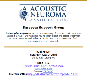
Dr. Danner will be speaking with the ANA Sarasota support group on April 7th, 2018. Many benefits can be gained from a supportive group environment. Family, friends, caregivers and interested persons are encouraged to attend.
TOPICS:
● Acoustic Neuroma
Sound Advice for Hearing Rehabilitation
Presented by Christopher J. Danner, MD, FACS
Tampa Bay Hearing and Balance Center
● Caring, Sharing, Networking and Support
FOR ADDITIONAL INFORMATION OR TO RSVP, PLEASE CONTACT:
Kim Simington, Support Group Leader/Facilitator
Phone: 908-963-8800 or Email: kjsimington@msn.com
Melanie Hutchins, ANA Manager of Volunteer Programs
Phone: 770-205-8211 or Email: volunteers@ANAUSA.org
The Miracle in the Christmas Play
After hearty handshakes and friendly hugs with a lovely young couple and their daughter, we walked away thinking back about seven years earlier. When I first met them, they sat quietly, eyes staring forward, unanimated, and I presumed, sadly. I had heard that they were coming in. The six-month old baby girl with them seemed unusually quiet as well.
The story stared with a pretty typical ear infection but something changed. She had become obtunded, lethargic, but also irritable. An urgent trip to the pediatrician resulted in emergency hospitalization and a spinal tap.
Meningitis had only been a vague term, not a familiar entity, not a personal reality. Its dreaded complications were even more unknown. On two intravenous antibiotics, initial hope that their third child, a girl after two boys, would rapidly improve faded as her lethargy persisted.
Five days into treatment, the reason became apparent: the bacteria were resistant to the usual antibiotics. With a medication change, she turned the corner and her personality showed glimmers of her prior vivaciousness. Soon thereafter, auditory brainstem response testing confirmed a major complication: the baby was now deaf, no signs of any ability to hear, not electrical and not even to loud sound. And that’s what brought them to see me, and ear specialist.
My wife and I marveled at this now sever-year old blonde delight with untamable rolls of curly hair, and an energetic personality to match. My eyes misted as I envisioned her performance that evening. In the Christmas play in the midst of several similarly aged children, she had been Angel Gabriel, reciting her lines perfectly:
“Do not be afraid, for behold, I bring you good tidings of great joy…”
Maternal prenatal smoking and hearing loss among adolescents
Although smoking and secondhand smoke exposure are associated with sensory hearing loss in children and adults, the possible association between prenatal smoke exposure and hearing loss had not previously been investigated. More than 12% of US children experience prenatal exposure to nicotine. Hearing test data were examined for 964 adolescents aged 12 to 15 years from the National Health and Nutrition Examination Survey 2005-2006. All children had undergone standardized audiometric testing. Blood tests and self-reports were used to identify adolescents exposed to secondhand smoke or active smokers. Prenatal exposure was defined as an affirmative parental response to, “Did the biological mother smoke at any time while she was pregnant with the child?” Standard criteria were used to define hearing loss.
Study Results: Parental responses affirmed prenatal smoke exposure in 16.2% of 964 adolescents. Prenatal smoke exposure was associated with at least some degree of hearing loss in the high frequencies. As well, prenatal tobacco exposed children had a higher rate of single sided low-frequency sensory hearing loss.
Conclusions and Relevance. Prenatal smoke exposure is independently associated with higher pure-tone hearing thresholds and an almost 3-fold increase in the odds of unilateral low-frequency hearing loss among adolescents. These findings suggest that in utero exposure to tobacco smoke may be injurious to the auditory system.
References
Comparative Benefit of Stapes Surgery for Hearing Loss in Children and Teens, Some with Congenital non-mobile Stapes, Some with Disease Caused non-mobile Stapes
Of the three hearing bones, the one most likely to become “stuck” or “non-mobile” is the third one, the stapes bone. All three hearing bones oscillate in response to incoming sound. The stapes bone transmits that sound vibration to inner ear fluids where the vibrations stimulate inner ear sensory hair cells. On a fairly rare basis in some children, the third hearing bone, the stapes bone, is non-mobile from birth as a result of a genetic defect or developmental error sometime, probably, in the 2nd trimesterof gestation in the womb. Also fairly rare is that some older children and teens suffer from a disease called otosclerosis. In otosclerosis, the stapes bone becomes stuck, non-mobile, first starting as a minimal problem, and then getting progressively worse. The pattern of hearing loss is quite similar in both groups.
On CAT scan imaging, the birth defect problem may have minor abnormalities of the inner ear and somewhat misshapen first (malleus) and second (incus) hearing bones. Some CAT scans looking at the birth defect problem find no abnormalities. The otosclerosis problem is almost always visible on the CAT scan, differentiating from the congenital problem. This article set out to compare how these two groups of children and teens improved with surgery on the stapes bone.
The measurements were hearing tests and chart reviews to see what complications might have occurred. Forty-four pediatric ears were studied, 27 with a congenital stapes problem and 17 with otosclerosis. Those with the congenital defect came to surgery at an earlier age, as expected, than did the otosclerosis group. Thirty seven percent of the congenital group had minor other hearing bone malformations whereas none of the otosclerosis ears had hearing bone problems other than the non-mobile stapes. Those who had otosclerosis generally gained a little more hearing from surgery than did the congenital ears, probably because some of the congenital ears had more wrong than just the non-mobile stapes bone but both groups uniformly gained hearing in a “statistically significant” manner following surgery. There were no cases of deaf ear and inner ear function remained stable in all but one ear which was a bit worse. There were no cases of serious complications, in either group. Compared to other published medical literature, this article had quite similar outcomes. Another disease entity which can cause a non-mobile stapes is called “tympanosclerosis.” Published literature reviewing stapes surgery in ears with tympanosclerosis found hearing gains from surgery to be less than with the congenital problem and the otosclerosis reviewed in this article.
The authors’ conclusion: When performed by an experienced surgeon, stapes surgery is safe and effecive in children who have either problem, although hearing results are generally better with otosclerosis than with the congenital problem.
References
Advantages of hyaluronic acid fat graft myringoplasty over fat graft myringoplasty.
Perforations of the ear drum membrane in children are not rare and can be a side effect of having had middle ear ventilation tube surgery. When a perforation persists either from infection or an ear tube or both, the question is how best to repair such with the least risk, the least anesthesia, and achieving a sufficiently high success rate. Fat grafts have been used for this purpose for many years but do not have uniformly high success rates. Use of micro-perforated hyaluronic acid sheeting has not been effective, either. This study combined the sheeting with the fat and compared the healing probability to just using a small piece of fat by itself. It was used for perforations of the ear drum membrane that occupied no more than 25% of the tympanic membrane surface. Postoperative hearing outcomes were compared, as well.
Ninety-two children with small tympanic membrane perforations were included in the study. Fifty were operated using either the sheeting combined with a small piece of fat and 42 were operated with the simpler, older fat placement technique. Age at surgery ranged from 4 to 17 years (mean age, 11.52 years).
Results: Successful closure of the ear drum membrane was achieved in 90% of the combined sheeting/fat graft group and in only 57% of the plain fat graft group. The postoperative hearing was understandably better in the combined fat-sheeting group as well. The probability of success did not depend on how old the children were at the time of surgery. No postoperative complications were observed. The locationof the perforation in the ear drum membrane was not found to be a factor indicative of failure.
Conclusions: The success rate of combining the hyaluronic acid sheeting with a small piece of fat was superior to the old technique of using fat alone. The Canadian authors suggested that this procedure could conceivably be done in a physician’s office.
Our comments: The clear need for sterility and safe sedation make this procedure best if done at an ambulatory surgery center or at a hospital. In addition, this procedure has a success rate of about 90% and is limited to small holes in the ear drum membrane compared to 97-98% succcess rates for larger perforations approached more invasively. In other words, this procedure takes minimal time by comparison as a trade offfor slightly lower success rates with the advantage of less potential for scarring and its effects on hearing. This procedure, then, has a place for small, relatively uncomplicated perforations in ear drummembranes.
References
- Advantages of hyaluronic acid fat graft myringoplasty over fat graft myringoplasty., , Arch Otolaryngol Head Neck Surg, 2012 Oct, Volume 138, Issue 10, p.950-5, (2012)